A Guide to Understand Knee Joint with Diagrams
The knee is the largest joint, and its primary function is helping in mobility. To understand the function and structure of the knee joint, a knee anatomy can be helpful. This article will introduce knee joint with knee anatomy.
1. Knee Joint Overview
The knee joint is a complex hinge joint. It is present at the lower part of the human body and functions to allow a back and forth motion without many side-to-side movements. Learning about knee anatomy can help the students to know about the structure and function of the knee.
To learn about the knees properly, the students can use knee anatomy diagrams. As drawing a knee joint by hand can be difficult and time-consuming, the students must use the EdrawMax Online tool, which can help them create a high-quality knee anatomy diagram.

2. The Structure of Knee Joint
There are totally 3 parts of knee joint, including ligaments, tendons, bones of the knee.
Ligaments of the knee:
Knees to be stronger as they hold a large portion of the body weight. There are four ligaments in the knee that connect one bone with the other. They provide strength to the knees.
- Anterior cruciate ligament and Posterior cruciate ligament: They are present in the knee joint, forming an "X" structure. Their function is to control the back and forth movement of the knee.
- Medial Collateral ligament and Lateral Collateral ligament: The collateral ligaments are present at the sides of the knee, and their primary function is to control the sideways motion of the knee.
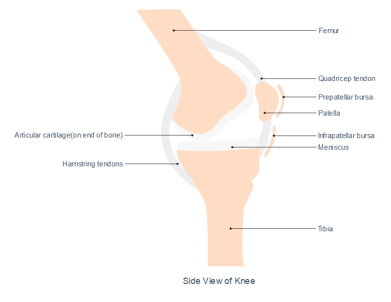
- Bursae of the knee: These are 14 bursae in total in the knee region. Their central function is to cushion the frictional force. They are of two types, communicating bursae and non-communicating bursae. The five main bursae are, prepatellar bursae, infrapatellar bursae, suprapatellar bursa, Pes Anserine Bursa, and semimembranosus bursa.
Tendons of the knee:
The tendons present in the knee are strong tissue bands that join the bones to the muscles. They give strength and stability to the joint. There is the quadriceps tendon, which attaches the thigh to the patella.
The patellar tendon connects the kneecap to the tibia.
Muscle of the knee: Three muscle groups provide strength to the knees.
- Quadriceps femoris muscle: They cross through the patella and work to extend the legs.
- Hamstring muscles: These muscles work to extend the hips.
- Popliteus muscles: They are present at the back of the knee and control the flexion of the knee. It also functions to unlock the knee by the rotation of the femur on the tibia.
Bones of the Knee:
The knee joint is a complex joint and is the meeting point of the femur and tibia. Fibula or calf bone is located at the lower legs and does not get affected by the action of the hinge joint. The patella bone is present at the center of the knee.
The medial and lateral meniscus acts as shock absorbers. They are disc-shaped and present between the femur and tibia to decrease the frictional force.
3. How to Create Knee Joint Drawing
There are mainly two ways to create the knee joint diagram, one is by hand, and the other one is using the easiest diagramming tool EdrawMax Online.
3.1 How to Create Knee Joint Drawing from Sketch
The students can make a knee joint diagram by hand, and for that, they need to follow these steps:
Step 1: >While drawing the knee joint, the students need to make a lump of bone at the end of the tibia. There is another lump at the end of the femur bone at the joint part. It sits on top of the tibia.
Step 2: For drawing the bones present in the knee joint, the students must include four of them. The femur is present at the hip and extended to the knee, while the tibia is present from knee to foot. Patella is located at the knee joint, and the fibula stays at the outer side of the legs.
3.2 How to Create Knee Joint Drawing Online
Creating a knee anatomy diagram by hand can be challenging. Hence, the students must use the EdrawMax Online tool. They need to follow simple steps to create a high-quality knee anatomy diagram.
Step 1: EdrawMax Online tool is user-friendly. Hence anyone can use this tool without any prior experience. To start with the work, the students need to open the EdrawMax Online and then open New. Under this part, there comes the Science and Education or Science Illustration option.
 Source:EdrawMax Online
Step 2: A user can use this tool for creating multiple sciences and education-related diagrams. They need to choose the Human Anatomy option present in this tab and look for the knee joint diagram.

Step 3: The tool allows a user to get a high-quality knee anatomy diagram without much hassle. After selecting a template, the students can modify it as per their requirements.
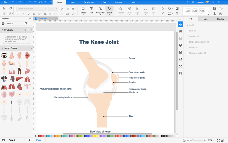
Step 4: Once the students are satisfied with their diagrams, they can save them in multiple formats and export them for their projects and dissertation papers. Or, even you can use the presentation mode once you finished your diagrams.

4. Types of Common Knee Injuries
In this part, here illustrates the types of common knee pain and injuries, and what caused the knee pain indeed.
Types of knee pain
Primarily, there are three categorizations for knee pain. They are pain due to acute injury, pain caused by medical conditions, and pains from overuse. Here are a few medical conditions which result in knee pain:
- Osteoarthritis
- Gout
- Osgood-Schlatter disease
- Popliteal cyst
- Cellulitis
- Septic Bursitis
- Osteomyelitis
Cause of knee injuries
As knee joints are highly used, and hence there are chances for it to get injured. Here are a few common causes of knee injuries:
- Sprains and strains or ligament injuries.
- Meniscus tear
- Fracture
- Bursitis
- Tendinitis
- Tendinosis
- Plica Syndrome
- Patellofemoral Pain Syndrome
5. Treatment Options for Knee Injuries
Treatment of knee injuries: The knee joint is a complex joint, and hence it requires medical attention if an individual injured their knee to avoid any severe circumstances. The conventional treatments of knee injury may include ice, physiotherapy, or pain reliever.
If an injury is minor, the person can continue with light exercises after 24-48 hours following the doctor's advice. Proper resting and not putting much weight on the knee are necessary to avoid further injury. The doctor may also prescribe the use of crutches.
In case of some serious knee injuries like an ACL tear, the patient may need arthroscopic surgery. Rehabilitation is pretty essential in this case. They may take the doctor's suggestions before starting physical therapy and exercises guided by professionals.
Injury prevention: An individual can prevent knee injuries, and for that, they need to do a few things:
- Maintaining a healthy weight not to pressurize the knees.
- Warming up before any intense sports and using knee guards when there are chances of injuries during sports.
- Doing hamstring exercises and weight training to strengthen the knees.
- Avoiding high-intensity workouts without any prior experience.
- Removing worn-out shoes
- Using seat belts to avoid hitting the knees.

6. Conclusion
The knee joint is a complex joint and plays a significant role in carrying the body weight and helps in walking. Hence, people should avoid knee injuries at any cost. A person can do exercises to strengthen their knees and prevent any high-intensity task or workout which may hurt their knees. In case they have an injury to the knee, they must seek medical attention without neglecting it.
To learn about knee anatomy, the students must use diagrams that can help them understand the structure and function of this joint. They must use the EdrawMax Online tool for creating a high-quality knee diagram.
In conclusion, EdrawMax Online is a quick-start diagramming tool, which is easier to make artery and vein diagram and any 280 types of diagrams. Also, it contains substantial built-in templates that you can use for free, or share your science diagrams with others in our template community.


