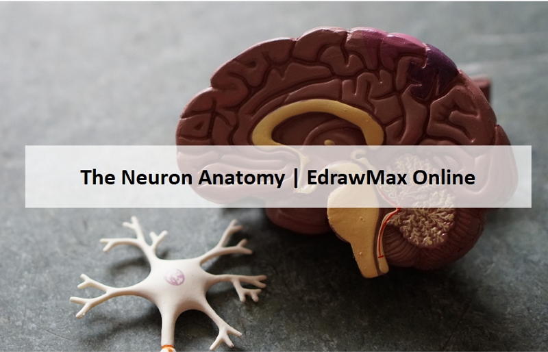
A Guide to Understand Neuron with Neuron Diagram
As the structural and functional unit of the nervous system, the neurons play a significant role in building communication between the brain and the body. This article introduces how to understand neurons and the functions with neurons labeled diagrams.
1. The Anatomy of a Neuron
Neurons are a significant part of the nervous system. They work as a communicator between the brain and the body. The neurons and glial cells make the brain. Neurons are cells that work to transmit the signal. They gather the stimuli for the command of action on which a human body works.
They carry the electrical or chemical signal to the central nervous system, where the data is analyzed. To properly understand the coordination between the brain and the body, the students must learn about the neurons. They can use neuron-labeled diagrams while learning the complex structure of neurons. Creating a neuron-labeled image by hand can be difficult. The students must use the EdrawMax Online tool to make a high-quality neuron diagram.
2. What Does a Neuron Look Like
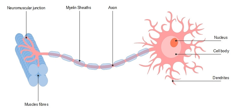
The main structure of a neuron includes the following parts:
- Dendrite: The cell body may have some branch-like structures, which work in receiving the signals. They are known as dendrite. A neuron may have multiple dendrites, while some of them may not have any. The dendrites receive signals from other neurons and pass them on to the cell body.
- Cell Body: The soma or cell body of the neurons has a nucleus, lysosomes, endoplasmic reticulum, and some other organelles. It has a membrane for protection and interaction with its surroundings. The functions of the cell body are maintaining the neuron’s formation, conveying the signal for the activities, and carrying the genetic information.
- Axon: The cell body is connected with the long tube-like structure, called Axon with Axon hillock. A neuron generally has a single Axon covered with insulations called Myelin sheath. The Axons convey the impulses to another neuron, and the myelin helps in conducting the signals.
- Synapse Vasculature: Medulla, present above the spinal cord, regulates the autonomic nerve outflow to the heart and blood vessels. The sympathetic nerves come out of the medulla to the spinal cord. Then there is a synapse with short preganglionic fibers, which creates a connection with sympathetic ganglia. The postganglionic efferent fibers, creating synapses in new places. They are forming it after coming to the heart and vasculature from the ganglia.
- Nerve Supply: The nervous system has two main sections, sympathetic and parasympathetic. The nervous system receives the stimuli acting information on the outside or inside the human body and then responds to it through actions. While the sympathetic nerves prepare the body for emergencies, the parasympathetic nerves maintain control for the ordinary time. Acetylcholine and norepinephrine work as neurotransmitters in the nervous system.
 Source: EdrawMax Online
3. How to Draw a Neuron Diagram
To learn about the structure of the neurons, the students can use a neuron labeled diagram. The students may follow these steps to make their neuron diagram, but the process is complex:
3.1 How to Draw a Neuron Diagram from Sketch
Step 1: First, the students need to draw a circle. Based on it, they need to draw a star-like shape. It is called the cell body of the neurons. One corner of the stars is extended, forming a very thin-tube-like structure–the Axon.
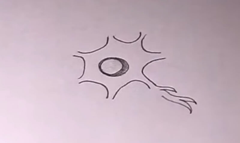
Step 2: The end part of the narrow tube is branched and has pretty small circles at the end. The branches are Axon terminals, while the ball-like structures are synapses. Next, the students need to create bulbs on the thin tube-like structure. They are known as Myelin Sheath.
Step 3: The corners of the star shape are extended, and the students need to draw irregular branches on them. These forms are known as dendrites. Finally, they have to make a circle in the star-like structure and put dots inside them. It makes the nucleus.

3.2 How to Draw a Neuron Diagram Online
However, creating a neuron labeled diagram by hand can be difficult, and the students may not find it good enough to use for their projects. To avoid such conditions, they must use the EdrawMax Online tool. The tool is user-friendly and can help to make a high-quality neuron labeled diagram. Here are a few simple steps which they have to follow:
Step 1: EdrawMax Online tool comes with an easy-to-use interface. It has helped it to gain a large number of users. Anyone can work comfortably on this tool which has made it a favorite diagramming tool of many users. To start the work, the students need to open the EdrawMax Online tool and then open New. Under New, they can look for the Science Illustration option.

Step 2: They can use the tool for creating multiple diagrams on science and education. It can help them to learn complex concepts. In this section, they have to find the Human Anatomy option. The students can find the neuron labeled diagram in this option.

Step 3:Once the students have selected their diagrams, they can smoothly work on them. They can modify the diagram according to their choice. It can help them to get a high-quality neuron labeled diagram apt for their projects and dissertation papers.
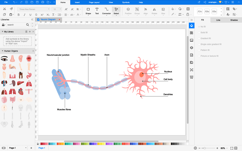
Step 4: Once the student completes their diagrams, they can save them in multiple formats. They can also export it and use it in their projects, papers, and studies. Or, it supports to use presentation mode for showing the science diagrams immediately.

4. The Classes of Neurons
Considering the function of the neurons, they are categorized in sections:
- Sensory Neurons
- Motor Neurons
- Interneurons
Sensory neurons work as the carrier of information. It gathers information about external or internal stimuli of the human body and carries it to the CNS. Here the data processing takes place. It is also related to the reflex action as they convey the electrical impulses to a relay neuron.
The motor neurons act to receive information about the stimuli from the other neurons and then accordingly signals the muscles and organs to take action. Its central function is to send impulses from the spinal cord to skeletal and smooth muscles to control muscle movements.
These neurons are present in the central Nervous system and act as the connector between the neurons. They take information about the stimuli from one neuron and then convey it to another. They work in both simple and complex circuits of the human brain.
5. Conclusion
As the fundamental units of nervous systems, they work to receive information about the stimuli. They also command the muscles in the human body. As neurons have complex structures, the students may use neuron-labeled diagrams to learn their structure and function. However, creating a neuron diagram by hand can be very difficult. The students must use the EdrawMax Online tool, which can help them create a good quality neuron labeled diagram for their lessons.
In conclusion, EdrawMax Online is a quick-start diagramming tool, which is easier to make artery and vein diagram and any 280 types of diagrams. Also, it contains substantial built-in templates that you can use for free, or share your science diagrams with others in our template community.