The Anatomy of Human Eye with Diagram
Create a Eye Diagram Online Free Free Download Free Download Free Download Free Download1. The Anatomy of Human Eye
The most complex sensory organs of the human body are the eyes. Every part of the human body is responsible for a specific action, from the muscles and tissues to the nerves and the blood vessels.
The human eye consists of many muscles and tissues that join to form an approximately spherical structure. It's primarily responsible for vision, color recognition, and the regulation of the human body's biological clock.
We may equate the human eye to a camera as each works by collecting, concentrating, and transmitting the light through the lens to produce an object image. The human eye diagram is a visual depiction of the human eye. The following aspects are essential when constructing a human eye diagram.
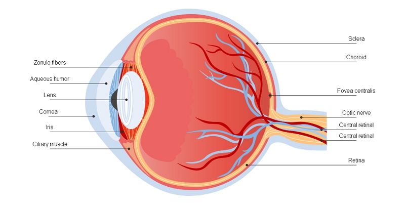
1.1 Conjunctiva of the Eye
The conjunctiva is a thin, translucent layer of tissue that protects the front of the eyes, including the sclera and the eyelids inner surface. Bacteria and foreign substances are stopped from going behind the eye by the conjunctiva.
The conjunctiva 's prime functions are:
- To make sure the front surface of the eye is moist and lubricated;
- Keep the eyelids' inner surface moist and lubricated so that they freely open and close without friction or discomfort of the eyes.
1.2 The Sclera of The Eye
The sclera is the eyeball 's thick connective tissue, which forms the eye's "white." The stroma layer of the cornea is continuous. The intersection of white sclera with the transparent cornea is called the limbus.
The sclera is the thick connective tissue of the eyeball, which forms the "white" eye. Furthermore, the sclera ranges in thickness from about 0.3 mm to 1.0 mm. It is made up of collagen (small fibers), arranged in irregular bundles and interlacing. The functions of Sclera:
- The eyeball shape is preserved by the sclera, along with the eye's intraocular pressure;
- The sclera's robust and fibrous structure also protects the eye from a severe injury from external injuries, such as laceration or rupture;
- The sclera also provides the extraocular muscles with a sturdy attachment, which controls eye movement.
1.3 The Cornea of The Eye
The cornea is the transparent front of the eye. It allows the light to enter the eyes, and it lies directly in front of the iris and the Pupil. When seen from the front of the eye, the cornea appears slightly broader than it is tall. That is because the sclera somewhat overlaps the top and bottom of the anterior cornea.
- The cornea not only allows light to enter the eye but also amounts to 65-70% of the eye's focusing power;
- Apart from allowing light to reach the eye, it is also responsible for the human eye's focusing power.
1.4 Iris of The Eye
The iris, the colored portion of the eye, regulates the amount of light entering the eye. The iris is a ring-shaped tissue with a central aperture, called the Pupil.
Iris consists of Ciliary body and Choroid. As for the Ciliary body, it covers the iris and is not apparent because it lies behind the opaque sclera. Yet, the Choroid is sandwiched between the eyeball 's rough outer sclera and the retina at the back of the eye.
Besides giving the eye its hue, the iris functions like a camera's diaphragm, which controls the Pupil's size. One muscle inside the iris constricts the Pupil in bright light, for example, full sunlight, and another iris muscle dilates the Pupil in dim lighting and darkness.
1.5 Pupil of The Eye
The pupil is in the middle of the iris opening. The pupil's purpose is to allow light to reach the eye so that it can concentrate on the retina to start the sight process.
- The iris and pupil together monitor how much light gets into the eye. Using a camera analogy, the pupil is the eye aperture and the iris is the diaphragm that controls the aperture size;
- Muscles within the iris monitor decide the size of the Pupil. Within the iris, the mechanism of muscle movement determines how much light the Pupil reaches the eye.
1.6 The Retina of The Eye
The retina is the sensory membrane, which forms the eyeball's inner surface. It is composed of many layers, including one that includes specialized cells known as photoreceptors. In the human eye, there are two types of photoreceptor cells, rods and cones.
- Photoreceptor cells receive corneal and lens-focused light and transform it into chemical and nervous signals transmitted through the optic nerve to visual centers in the brain;
- These signals are interpreted into representations and visual images in the brain's visual cortex.
1.7 Optic Nerve of The Eye
A bundle of over 1 million nerve fibers, the optic nerve is responsible for the transmission of nerve impulses from the eye to the brain. Those nerve signals contain brain processing information. The optic nerve's front part, visible on the retina, is called the optic disk, or optic nerve head.
- The optic nerve transmits sensory information, including the perception of light, perception of color, and contrast;
- It also carries out the sensory impulses responsible for two crucial neurological reflexes, the reflex of light, and the reflex of accommodation;
- The light reflex refers to the constriction that happens in both pupils when light shines through either eye.
2. Facts about The Human Eye
As the window of the heart, eyes contains some facts that people had few noticed.
- Blind people with eyes also can feel the difference between light and darkness. There are individual cells in the eyes which detect light but are not involved in image formation;
- Each eye has one tiny blind spot. It is the place where the eyeball is connected to the optic nerve. The loss in vision is not visible, because each eye fills in the blind spot of the other;
- Babies are born with eyes of all sizes. From birth until death, human eyes stay around the same size.

3. Key Takeaways
The human eye enables vision as a conscious sense organ; the rod and cone cells of the retina allow for conscious perception and vision of light, including color distinction and depth perception. The human eye can discern nearly 10 million colors.
EdrawMax is a wondrous full-featured application commonly adopted for making the human eye diagram. Now users will create a perfect graph in moments with full impact with only a few clicks. It is extremely user friendly and offers 100% privacy when it comes to security.