A Guide to Understand Animal Cell with Diagram
Cells are the structural and functional unit of the body, and hence to understand the structure and function of an animal body, it is crucial to learn about the animal cells. There are multiple organelles and nuclei, which makes learning complicated. The students can use diagrams for a better understanding. It can be challenging to draw an animal cell diagram by hand, and hence the students must use the EdrawMax tool.
1. What is the Animal Cell?
The Animal cells are the structural and functional units of animal bodies and are eukaryotic in nature. The animal bodies can be unicellular or multicellular. They are membrane-bound and have different organelles.
Shape and Size:
- The animal cells have different shapes and sizes. The largest animal cell is an ostrich egg with a 5-inch diameter, while the smallest animal cell is a neuron having a 100 microns diameter.
- As the animal cells are Eukaryotic, they have membrane-bound organelles. Most of them carry genetic material within the membrane-bound nucleus.
Functions:
- They give shapes to the cell, and they participate in the body's functions.
- They can produce energy and store it.
- They participate in fluid and molecule transportation.
2. The Structure of Animal Cells
Animal cells are eukaryotic cells, and they lack rigid cell walls. They have membrane-bound nuclei to carry the genetic material like DNA. The animal cells also have organelles that participate in the functions of the cells.
- Centrioles:
- When mitosis occurs, the centrioles replicate. After the division, when the cytoplasm is formed, the microtubules bind the chromosome into centromere.
- The microtubules of centrioles help in the transportation of different substances linked with the glycoprotein.
- Endoplasmic Reticulum:
- They have a large surface area which is the site for chemical reactions.
- They have enzymes that make them a place for lipid synthesis.
- Protein manufacturing, processing, and transportation for cell works.
- Endosomes:
- Folding of Plasma membrane and diffusion of molecules.
- Remove excretory materials from the cells by exocytosis and phagocytosis.
- Plasma Membrane:
- It protects the content of the cell.
- Regulates the process of homeostasis and allows selective liquids to pass through the cell membrane.
- Protein helps in the circulation of the materials within the cell.
- Nucleus:
- The primary work of the animal cell is cell growth and regulation of metabolism.
- It carries genetic information, and the nucleus acts as an information center.
- Acts as the site of Transcription.
- Cytoplasm:
- It gives shape to the cells.
- Holds the components and stores molecules that are essential for cellular processes.
- It protects the components of cells and organelles from damage.
- Mitochondria:
- Their central function is to generate ATP.
- They store calcium and participate in cell signaling.
- It plays a significant role in apoptosis or programmed cell death.
- Ribosomes:
- Acts as the site of protein synthesis.
- Acts as the site for the genetic coding of proteins.
- Golgi apparatus:
- They transfer and modify proteins and lipids and send them to the required sites.
- Lysosomes:
- Acts as the site where digestion of cell nutrients and excretion occurs.
- The enzymes help in breaking down proteins, carbohydrates, and lipids.
- Cytoskeleton:
- Organize the cell components and maintain the shape.
- Help in giving cells and their organelles a uniform movement.
- Microtubules:
- Provides structural support and organizes the components of the cytoskeleton.
- They participate in the formation of spindle fibers.
- The primary elements for the cilia-flagella creation.
- Peroxisomes:
- Lipid metabolism
- Chemical detoxification
- Cilia and Flagella:
- Flagella helps in sperm cell movements.
- Cilia assists the movement of fluid away from the immobile cells.
- Vacuoles:
- Storage of food, water and removes waste and toxic materials
- Acts as a guard when substances pass by it.
- Microvilli:
- The microvilli present in the small intestine increase surface area for absorption of food and water.
- The white blood cells behave like an anchor helping in free movement in the bloodstream.
They are found in animal cells and have nine microtubule bundles. They replicate themselves and participate in cell division.
Functions:
These organelles look like folded membranes and form a thin network of sacs that connects the cytoplasm and nucleus of the cell.
Functions:
They are vesicle-bound organelles found in the cytoplasm.
Functions:
It is a semi-permeable membrane made of protein and lipid present in all living cells. It creates a thin layer to form a separation between the cell and the external environment.
Functions:
The nucleus is a spherical-shaped organelle covered with a double-layered cell membrane and carries the genetic material in the DNA form. The nucleus stays on the cytoplasm with the help of filaments and microtubules. A cell generally has one nucleus, but it can be multinucleated as well.
Functions:
The cytoplasm is a gel-like structure present within the cell membrane and contains cell organelles. It has organelles like ribosomes, mitochondria, endoplasmic reticulum, Golgi apparatus, microtubules, etcetera.
Functions:
They are membrane-bound organelles present in the eukaryotic cells. They have double membranes, and the folds of the membrane are called cristae. They can be oval-shaped or maybe spherical and rod-shaped.
Functions:
Ribosomes are tiny organelles and present in all living cells and stay in the cytoplasm and endoplasmic reticulum. It consists of 60% of RNA cytoplasmic granules and 40% proteins.
Functions:
They are membrane-bound organelles held by the protein matrix. The animal cells have 1-2 Golgi Bodies, while the plant cells have hundreds of them.
Functions:
Lysosomes or cell vesicles are round subcellular organelles found in most eukaryotic cells.
Functions:
The cytoskeleton is a network made up of long protein chains and amino acids. They are present in the cytoplasm of eukaryotic cells.
Functions:
They are hollow cylindrical structures found throughout the cytoplasm of the animal cells.
Functions:
They are spherical shapes, membrane-bound organelles.
Functions:
They are the locomotive projections present on the cell surface.
Functions:
They are membrane-bound sacs found in the cytoplasm. They have a single membrane, namely the tonoplast.
Functions:
They are surface protrusions formed from the actin filament’s accessory protein and are primarily there in the intestinal lining and white blood cells.
Functions:
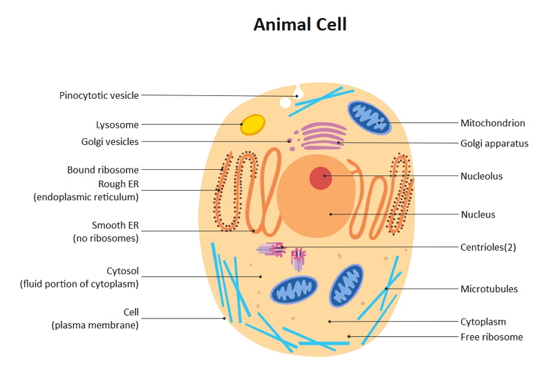 Source:EdrawMax Online
3. How to Draw the Animal Cell Diagram
As animal cells have different organelles, it is tough to draw an animal cell by hand. Here are a few steps that a student can follow to create an animal cell diagram:
3.1 How to Create Animal Cell Diagram from Sketch
Creating such a diagram by hand can be difficult. The students can follow these steps to make their elbow joint diagram:
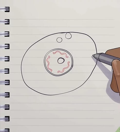
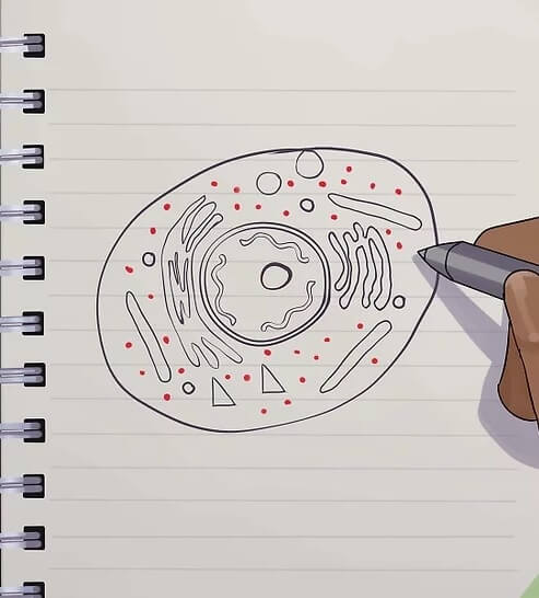
Step 1: Draw a large oval shape and then add one-two small circular figures within them.
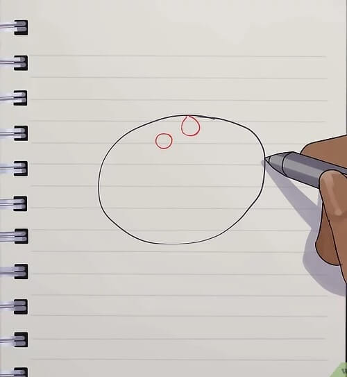
Step 2: Draw two concentric circles for the cell nucleus, and a pretty tiny gap between them. Create a shadowed circle within it for the nucleolus. Form squiggle structure within for chromatin fibers.
Step 3: Draw rod-shaped mitochondria, dumbbell-shaped Golgi apparatus, finger-like endoplasmic reticulum, circles for lysosomes, and small rectangles for centrioles. Once done, they need to add a few small circles for vacuoles and then put dots all over the cytoplasm for ribosomes.
3.2 How to Create Animal Cell Drawing Online
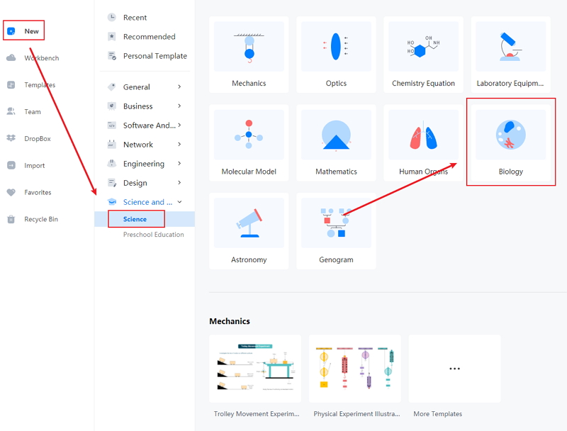
Since it is challenging to create an animal cell diagram by hand, the students must use the EdrawMax Online tool. It makes it easier to create a high-quality image without much hassle. For that, the students must follow these few simple steps:
Step 1: EdrawMax online is a user-friendly tool, and hence anyone can work on them without much experience. It allows the students to have a high-quality animal cell diagram. The students need to open the EdrawMax online tool and then open New. Then they have to click on the 'Science and Education' section.

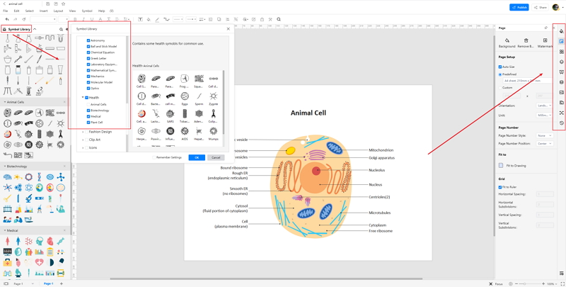
Step 2: The students can use the tool for creating multiple images. For making animal cell diagrams, the students need to select the 'Biology tab.' Now they should look for the animal cell diagram. The students can easily edit and modify the diagram to suit their requirements.
Step 3: After selecting the template they require, the students should modify it as per their choice. The tool gives the students some hassle-free options to edit their diagrams. They can work on those images according to their preference to create a high-quality animal cell image.
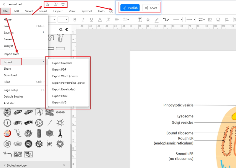
Step 4: Once they finish, they can easily save the file in multiple formats. They can use it for their studies or use it in their projects or dissertation papers.
4. Animal Cell VS. Plant Cell
Both animal cells and plant cells are eukaryotic and have membrane-bound organelles. However, there are some differences between the two types of cells:
- The plant cells have cell walls and cell membranes for rigidity. However, in animal cells, there is only one cell membrane that is present.
- Though both animal cells and plant cells have mitochondria, the chloroplast is only present in plant cells.
- Plant cells have large vacuoles, while animal cells have multiple and small vacuoles.
5. Conclusion
The animal cells form the body and give shape. They also participate in all the main functions of the body like metabolism, transportation of liquids. Therefore, to understand the body systems, it is significant to learn about animal cells. The students can use animal cell diagrams to create animal cells, and for that, they must use EdrawMax online tool, which can help them have a high-quality diagram.
In conclusion, EdrawMax Online is a quick-start diagramming tool, which is easier to make animal cells diagram and any 280 types of diagrams. Also, it contains substantial built-in templates that you can use for free, or share your science diagrams with others in our template community.
Related Articles